Clinic locations

Dental Radiology
Essential in Dental Diagnosis and Treatment Planning

What is dental radiology?
Dental radiology is a fundamental branch of modern dentistry, which uses medical imaging technology to obtain detailed images of tooth, bone and surrounding tissue structures. This allows dentists to identify hidden dental problems and plan appropriate treatments. Dental X-rays provide a clear view of internal structures such as tooth roots, jaw bones, temporomandibular joints, and the sinus cavity, thus allowing for accurate diagnosis and minimally invasive interventions.
Modern dental radiology has developed enormously in recent decades, moving from conventional film radiographs to advanced digital techniques such as CBCT (Cone Beam Computed Tomography) and digital panoramic radiographs, which provide high-resolution three-dimensional images. These advances have allowed for greater accuracy and efficiency in diagnosis and treatment, while reducing radiation exposure.
What does dental radiology do?
Dental radiology plays a key role in evaluating oral health, providing detailed information that cannot be obtained by visual examination of the oral cavity alone. This helps dentists detect problems in their early stages, before they become symptomatic or visible to the naked eye.
The main goals of dental radiology are:
- 1. Early detection of dental problems: Dental x-rays can reveal cavities hidden between teeth, under fillings, or below the gum line, which are not visible during a routine clinical exam.
- 2. Evaluation of jawbones and impacted teeth: Dental radiology is essential to evaluate the condition of the jawbones and to diagnose problems such as impacted teeth (teeth that have not fully erupted), cysts, tumors, or other abnormalities of bone structures.
- 3. Complex treatment planning: X-rays are used to plan various dental procedures, such as orthodontic treatments, dental implants, impacted tooth extractions, or periodontal surgeries.
- 4. Long-term dental health monitoring: Dental x-rays are used to track the progress of treatments and verify whether they have been effective. For example, x-rays are essential for monitoring root canal treatments, dental implants, and long-term prosthetic restorations.
- 5. Evaluation of periodontal disease: X-rays help assess the level of bone loss in periodontal disease, providing a clear picture of the progression of the disease and the need for further interventions.
Types of Dental X-rays
There are several types of dental x-rays, each with different purposes and used depending on the diagnostic and treatment needs. Depending on the complexity and location of the dental problems, the dentist may recommend one or more types of x-rays. Here are the most common types of dental x-rays:
Intraoral X-rays
Intraoral x-rays are the most commonly used and provide detailed images of the teeth, gums, and jawbone. They are performed by placing the X-ray film inside the oral cavity, and the radiation beam is directed from the outside.
- 1. Bitewing X-ray: This type of X-ray is used to evaluate interdental cavities (between the teeth), as well as to evaluate the bone structure around the teeth. Bitewing X-rays are performed by placing a X-ray film between the teeth, so that both the upper and lower tooth crowns are visible. They are essential for the early detection of cavities and periodontal disease.
- 2. Periapical X-ray: Provides a detailed image of the entire tooth, from the crown to the root, including the bone around the tooth. Periapical radiographs are used to diagnose dental infections, abscesses, cysts, as well as to evaluate the condition of the roots of teeth after endodontic treatments.
- 3. Occlusal radiography: This type of radiography is used to visualize the entiredental arch. Occlusal radiographs are useful for identifying teeth that have not erupted properly, bone fractures, or to evaluate the structure of the maxillary and mandibular bones. They are also used to detect the presence of foreign bodies in the oral cavity or to evaluate tooth development in children.
Extraoral radiographs
Unlike intraoral radiographs, extraoral radiographs provide an overview of the jaws, teeth, and facial structure. They are used to diagnose more complex problems or plan surgical treatments.
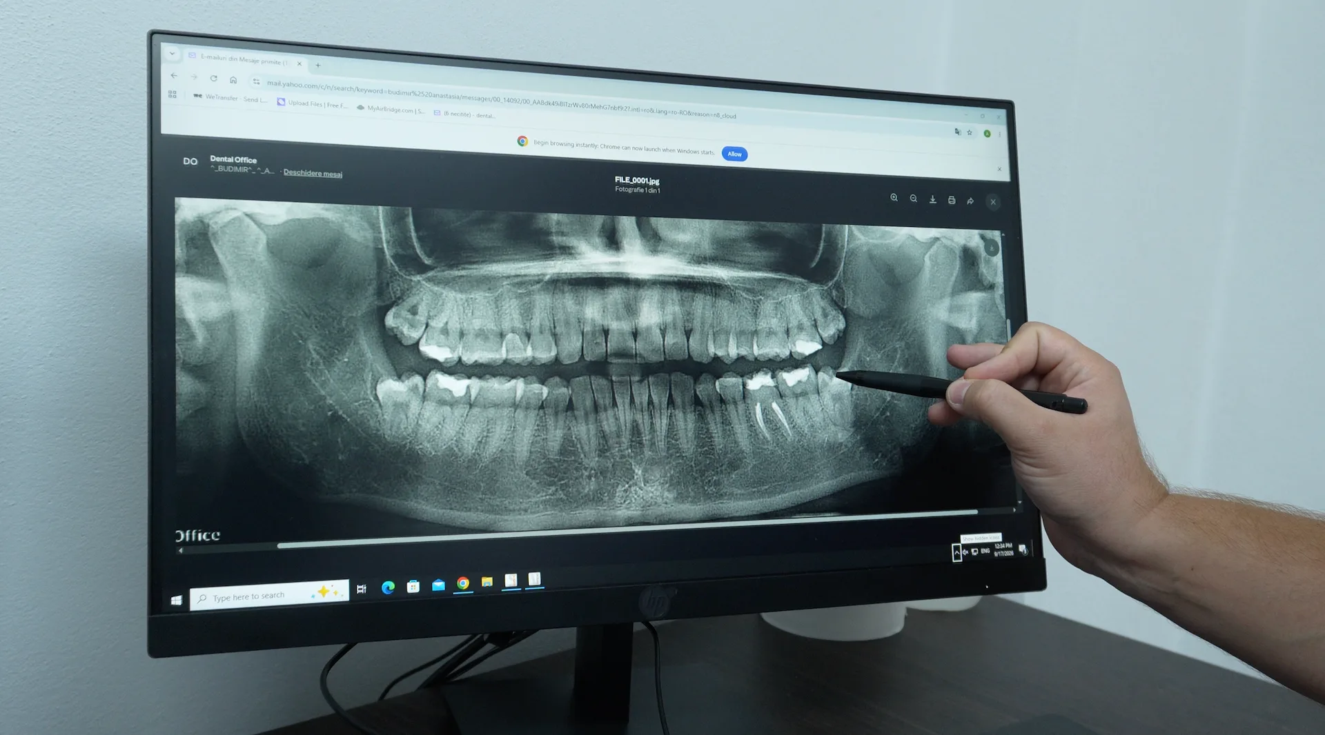
- 1. Panoramic radiograph (Orthopantomography – OPG): This provides a complete image of the entire oral cavity, including the teeth, jaw bones, temporomandibular joints, and sinus cavities. It is frequently used to assess the general state of oral health, to plan extractions of impacted teeth, dental implants or to detect bone abnormalities and infections. Panoramic radiography is an indispensable tool in orthodontics, implantology and oral surgery.
- 2. CBCT (Cone Beam Computed Tomography): CBCT is one of the most advanced dental imaging methods, providing highly detailed three-dimensional images of teeth, roots, jawbones and surrounding structures. This is essential for planning complex interventions such as dental implants, orthodontic treatments and orthognathic surgery. CBCT allows dentists to visualize anatomical structures with much greater precision than conventional radiographs.
- 3. Cephalometric radiography: This is a lateral radiograph of the skull that provides a clear image of the structure of the facial bones and teeth. It is commonly used in orthodontics to assess the relationships between the maxilla, mandible, and occlusion (bite). Cephalometric radiographs are essential for planning complex orthodontic treatments and monitoring progress during them.
- 4. Sialography: It is a specialized radiological technique that focuses on the salivary glands and their ducts. A contrast agent is used that is injected into the salivary glands so that they are clearly visualized on the radiograph. This is used to diagnose problems such as blockages or infections of the salivary glands.
Technology Used in Dental Radiology
Modern dental radiology relies on advanced technologies, designed to improve image quality and reduce radiation exposure. Technological advances have led to the development of digital radiographs, which provide clearer images with a much lower radiation dose than traditional film radiographs.
Key technologies used in dental radiology:
- 1. Digital radiography: Digital radiography has largely replaced traditional film radiography. Instead of using radiation-sensitive film, digital radiography uses electronic sensors that capture the dental image and transmit it directly to a computer monitor. This technology offers numerous advantages, including:
● Reduced radiation exposure: Digital radiography exposes patients to significantly less radiation compared to traditional film techniques. This is essential for patient protection, especially for those who require frequent X-rays, such as children or orthodontic patients.
● Superior image quality: Digital images are of much better quality, allowing for quick adjustments, such as magnification or changing the contrast, to facilitate interpretation and diagnosis. They can also be stored digitally, quickly shared with other specialists, and used for treatment planning.
● Faster processing time: Digital images are available instantly after the X-ray is taken, eliminating the need for film development time. This allows the dentist to provide an immediate diagnosis and plan treatment without delay. - 2. CBCT (Cone Beam Computed Tomography): CBCT is a three-dimensional imaging technology that has revolutionized dental radiology. This technology allows for detailed images of dental, bone and soft tissue structures, providing a precise and complete view.
● 3D Imaging: Unlike traditional two-dimensional radiographs, CBCT provides three-dimensional images of the jaw, teeth and surrounding structures. This 3D capability is essential for dental implants, because it gives the doctor a clear picture of the thickness and density of the bone, as well as the exact position of the nerves and sinuses.
● Highly accurate diagnosis: CBCT is used to diagnose complex conditions, such as fractures of the jaw bones, impacted teeth, or complicated occlusal relationships. It also allows for a detailed assessment of the temporomandibular joint and sinus cavities.
● Low radiation doses: CBCT uses a cone beam of radiation to obtain 3D images, which means that radiation exposure is much lower than with conventional computed tomography.
- 3. Digital Panoramic X-rays (Orthopantomography - OPG): OPG is a digital technology that provides a panoramic image of the oral cavity, including the teeth, jaw bones, temporomandibular joints, and sinuses. It is frequently used for general oral health assessment, planning extractions of impacted teeth, or for orthodontic and implantology planning.
● Complete jaw assessment: Panoramic radiography allows the doctor to see the entire dental and bone structure in a single image, making it ideal for diagnosing complex problems involving multiple areas of the oral cavity.
● Speed and efficiency: The procedure is fast and comfortable for the patient, while providing a detailed and accurate image.
The Role of Radiography in Dental Diagnosis and Treatment
Dental radiography is an indispensable tool for the diagnosis and treatment of dental conditions. Without radiological imaging, many dental problems may go undiagnosed until symptoms become apparent, often when the condition is at an advanced stage and difficult to treat. Dental radiography allows for early detection and rapid intervention, preventing serious complications and ensuring less invasive and more effective treatment.
How does radiography help diagnose dental conditions?
- 1. Detection of hidden cavities: Dental radiographs are essential for identifying cavities that are not visible on a clinical examination, especially interdental cavities and those under old fillings. In many cases, these cavities are detected at an early stage, allowing for rapid and minimally invasive treatments, such as fillings.
- 2. Diagnosis of dental infections and abscesses: Infections at the tooth root or in the bone can be detected with the help of periapical radiographs. These images allow doctors to assess the extent of the infection and decide on the appropriate treatment, whether it is a root canal (endodontic) or surgery.
- 3. Assessing periodontal disease: Dental x-rays are crucial in assessing the stage of periodontal disease, especially in terms of bone loss. Periapical or panoramic x-rays can show how much bone has been affected by chronic inflammation and help monitor the progression of the disease and the effectiveness of periodontal treatments.
- 4. Identifying impacted or supernumerary teeth: Panoramic x-rays are essential for identifying teeth that have not erupted properly, such as wisdom teeth, or other dental anomalies, such as supernumerary teeth. They provide a clear picture of the position of the teeth and help plan for extractions or other necessary orthodontic treatments.
- 5. Detection of bone lesions and cysts: X-rays are often used to diagnose cysts, tumors, or other lesions of the jawbone, which can affect the overall health of the oral cavity. In some cases, these lesions may be asymptomatic and can only be identified through radiological imaging.
Importance of X-rays in Dental Treatment Planning
Patient Safety and Protection in Dental Radiology
One of the common concerns related to dental radiology is radiation exposure, but modern technologies have significantly reduced this risk.
Safety Measures in Dental Radiology
In modern dental radiology, patient safety is an absolute priority. Even though dental X-rays involve radiation exposure, the amount of radiation emitted is extremely small, especially when digital technology is used. However, to further minimize the risk, a series of protective measures are implemented.
- 1. Reducing radiation exposure: One of the most important improvements in modern dental radiology is the significant reduction in radiation exposure to patients. Digital radiographs require up to 80% less radiation than traditional film radiographs, making this technology much safer for patients.
- 2. Use of protective equipment: To provide additional protection, patients wear lead aprons and thyroid collars during radiological procedures. These help protect sensitive organs, such as the thyroid gland, from unnecessary radiation. Also, especially in the case of children and pregnant women, dentists take additional measures to minimize exposure.
- 3. Adherence to the ALARA principle: In dental radiology, the ALARA principle (As Low As Reasonably Achievable) is an important guideline for reducing radiation exposure. Dentists use X-rays only when absolutely necessary and optimize the settings of the machines to ensure the lowest possible dose of radiation without compromising image quality.
- 4. Strict protocols for radiological equipment: Radiological equipment in dental offices undergoes regular inspections to verify proper functioning and to ensure that radiation is emitted only at the desired level. Modern X-ray machines are also equipped with precise timers that ensure that the radiation beam is exposed for the shortest possible time.
Addressing common radiation concerns
Even though radiation exposure during dental X-rays is very low, patients often have questions and concerns about the safety of these procedures. It is important for dentists to provide clear information and address these concerns to reassure patients that the risks are minimal.
- 1. Radiation from dental x-rays vs. natural radiation: To provide a clearer perspective, the radiation exposure from a dental x-ray is often compared to the natural radiation we are exposed to every day. For example, a routine digital x-ray involves radiation exposure equivalent to several hours spent outside, exposed to cosmic or natural radiation from the ground.
- 2. Frequency of dental x-rays: Patients often wonder how often they should have dental x-rays. The answer depends on their oral health and their risk of developing dental disease. In general, patients with good oral health may need x-rays every 1-2 years, while those with chronic dental conditions or an increased risk of cavities may need x-rays more frequently.
- 3. X-rays in children: Children may need dental x-rays more frequently than adults because their teeth and jaws are still developing. X-rays are essential for monitoring the development of permanent teeth and for early detection of potential problems, such as impacted teeth or malocclusions.
- 4. X-rays during pregnancy: Although it is recommended to avoid radiation exposure during the first trimester of pregnancy, dental X-rays are safe during the rest of the period if absolutely necessary and appropriate protective measures, such as a lead apron, are used.
Patient Preparation for X-ray
Dental X-rays are simple and quick procedures, but It is important that patients are informed about what the preparation and procedure involve. Good communication between the doctor and the patient helps reduce anxiety and ensures a procedure without complications.
What should patients know before a dental x-ray?
- 1. Understanding the need for the procedure: Before the x-ray, the dentist will explain to the patient why it is necessary and what information it provides. The doctor will also discuss the risks and answer any questions related to radiation exposure.
- 2. Preparing for the procedure: In the case of intraoral x-rays, the patient may be asked to remove any jewelry, glasses, or metal objects that could interfere with the radiological image. Also, for additional protection, the patient will wear a lead apron that covers the chest and abdomen, as well as a collar to protect the thyroid gland.
- 3. Indications for patients with dentures or orthodontic appliances: Patients who wear removable dentures or orthodontic appliances should be informed that some of these may be temporarily removed before the x-ray is taken, so as not to affect the quality of the image.
- 4. Patient comfort: The procedure is usually quick and painless, and the dentist or radiologist assistant will provide all the necessary instructions to make the patient feel comfortable. During intraoral x-rays, the patient will need to keep the radiological film or digital sensor in position, which may cause slight discomfort in some cases, but this is temporary.
Dental radiology is an essential pillar in the diagnosis and treatment of dental conditions, providing a detailed view of the internal structures of the oral cavity. Whether it's early detection of cavities, assessment of bone loss in periodontitis, planning dental implants or diagnosing complex conditions, dental X-rays are indispensable in modern dentistry.
With the help of advanced technologies such as digital radiography and CBCT, patients benefit from high-quality images with minimal radiation exposure. Also, additional protective measures such as lead aprons and compliance with the ALARA principle ensure the safety of patients and medical personnel.
Dental X-rays not only help in the early detection of dental problems, but also play an essential role in planning complex treatments such as dental implantology, orthodontic treatment and dental surgery. Radiology provides a clear and detailed image of the oral cavity, allowing dentists to plan and execute personalized treatments tailored to each patient's needs.
Dental radiographs also contribute to the long-term monitoring of dental treatments. They help doctors evaluate the success of treatment, identify potential complications, and take corrective measures when necessary.
By addressing patient safety and protection, modern dental radiology ensures that patients are exposed to minimal doses of radiation, without compromising the quality of diagnosis and treatment. Patient preparation, whether it is removing temporary dentures or understanding the procedure, plays an important role in ensuring the success of the x-ray and in effective collaboration between the doctor and the patient.
In short, dental radiology is an essential tool in modern dentistry, allowing doctors to provide accurate, safe and effective treatment, directly contributing to the oral health and overall well-being of patients.
Frequently Asked Questions (FAQ) about Radiology
-
1. What is dental radiology and how does it help diagnose dental problems?
Dental radiology is an imaging technology used to obtain detailed images of the teeth, jawbones, and soft tissues in the oral cavity. It helps dentists detect hidden cavities, infections, cysts, bone loss, or other abnormalities that cannot be seen with a simple clinical exam.
-
2. What are the main types of dental x-rays available?
The most common types of dental x-rays include bitewing, periapical, panoramic (OPG), and CBCT (Cortex Computed Tomography) x-rays. Conical). Each type provides detailed images and is used to diagnose different dental conditions or plan complex treatments.
-
3. How often should I have a dental X-ray?
The frequency of dental X-rays depends on the oral health of each patient. People with good oral health can have X-rays every 1-2 years, while patients with dental conditions, such as frequent cavities or periodontal disease, may require more frequent X-rays, as recommended by their dentist.
-
4. Are dental X-rays safe? What is the level of radiation exposure?
Modern dental X-rays, especially digital ones, expose patients to a very low dose of radiation. In addition, protective measures, such as lead aprons, are used to further minimize exposure. The amount of radiation is comparable to what we are naturally exposed to on a daily basis.
-
5. What is CBCT and why is it used in dentistry?
CBCT (Cone Beam Computed Tomography) is an advanced 3D imaging technology that provides a detailed view of the teeth, jaws and soft tissues. It is used for planning dental implants, evaluating impacted teeth and diagnosing complex conditions, offering much greater accuracy than conventional x-rays.
-
6. How long does a dental x-ray take and is it painful?
Dental x-rays are quick procedures, taking just a few minutes. They are completely painless and only involve maintaining a comfortable position while the image is being captured. Results are available immediately with digital X-rays.
-
7. Do I need to prepare in any way for a dental X-ray?
Preparation for a dental X-ray is minimal. The patient may be asked to remove jewelry or removable dentures, if applicable, and will wear a lead apron for protection. Otherwise, the radiography is performed quickly and without discomfort.